163 results
- Digital Images
- Online
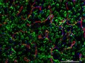
Cellular architecture of normal human skin imaged by whole mount tissue microscopy. Human skin has a rich network of white blood cells (specifically dendritic cells, T cells and macrophages) which form sheaths around blood vessels. This image was taken directly beneath the junction that joins the dermal and epidermal layers of the skin (dermo-epidermal junction). At this level, the capillary network (stained for CD31; red) is visualised against a lawn of autofluorescent dermal papillae (finger-like projections of the dermis; green) scattered with dendritic cells (stained for CD11c; green) and macrophages (stained for LYVE-1; blue). This normal cellular architecture is grossly disrupted in diseased skin (see related images). Scale bar (white) represents 200 micrometres.
Dr. Xiao-nong Wang, Human Dendritic Cell Laboratory, Newcastle University- Videos
Cell. Part 2, The chemistry of life.
Date: 2009
- Digital Images
- Online
Plasma cell, lymphocyte and fibroblast
Rob Young
- Digital Images
- Online
Human T cells showing nuclei
A. Walker, L. Sharp & J. Pryde
- Books
- Online
Leukosin : a new substance found in the blood of leukamia : also a description of another crystalline body found in the vomitus / by James C. White.
White, James C. (James Clarke), 1833-1916.Date: [1859?]
- Books
- Online
On the identity of the white corpuscles of the blood with the salivary, pus, and mucous corpuscles / by Joseph G. Richardson.
Richardson, Joseph Gibbons, 1836-1886.Date: [between 1860 and 1869?]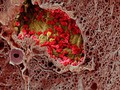
- Digital Images
- Online
Blood vessel in a melanoma, SEM
S. Gschmeissner, K. Hodivala-Dilke & M. Stone
- Digital Images
- Online
Skin around wound, mouse, blood cells
David Gregory & Debbie Marshall
- Digital Images
- Online
Thyroid nodule smear showing hematoidin crystals
William R. Geddie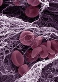
- Digital Images
- Online
Blood clot
Kevin Mackenzie, University of Aberdeen
- Digital Images
- Online
SEM of red blood corpuscles, close-up
David Gregory & Debbie Marshall
- Digital Images
- Online
SEM of blood clot + one white corpuscle
David Gregory & Debbie Marshall- Pictures
Shigellosis: acute: microscopic view of macrophage cells (white blood cells). Drawing by P.H. Manson-Bahr, ca. 1930.
Manson-Bahr, Philip H. (Philip Henry), Sir, 1881-1966.Date: 1930Reference: 571049i
- Digital Images
- Online
SEM of blood clot + one white corpuscle.
David Gregory & Debbie Marshall
- Digital Images
- Online
Glomerular capillaries and podocyte
University of Edinburgh
- Digital Images
- Online
Normal spleen showing B cells and T cells
Peter Lane and Fiona McConnell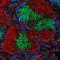
- Digital Images
- Online
Normal spleen showing B cells and T cells
Peter Lane and Fiona McConnell
- Digital Images
- Online
Normal spleen showing B cells and T cells
Peter Lane and Fiona McConnell
- Digital Images
- Online
Normal spleen showing B cells and T cells
Peter Lane and Fiona McConnell
- Digital Images
- Online
Normal spleen showing B cells and T cells
Peter Lane and Fiona McConnell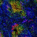
- Digital Images
- Online
Normal spleen showing B cells and T cells
Peter Lane and Fiona McConnell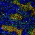
- Digital Images
- Online
Normal spleen showing B cells and T cells
Peter Lane and Fiona McConnell
- Digital Images
- Online
Polymorphonuclear leucocyte - TEM
Rob Young
- Digital Images
- Online
Polymorphonuclear leucocytes - TEM
Rob Young
- Digital Images
- Online
Fungal elements engulfed by neutrophils
Rob Young